Medical diagnostics relies on the analysis of clinical data such as medical images, laboratory measurements, physiological signals, and patient records to identify disease. In recent years, Artificial Intelligence (AI), particularly machine learning and deep learning methods, has been applied to these data sources to assist with diagnostic tasks. Algorithms trained on labeled datasets can classify medical images, detect abnormalities, and identify statistical patterns associated with specific conditions. These computational approaches are being studied and deployed in areas such as radiology, pathology, and clinical decision support, where large volumes of data must be interpreted as part of the diagnostic process.
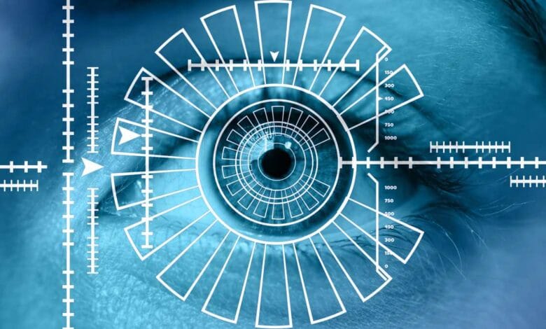
Scientists have found a way to learn about lung and heart problems in premature babies by examining pictures of their eyes. The blood vessels in the back of the eye can provide information about the rest of the body. With artificial intelligence, doctors can detect signs of complications earlier. Retinal photographs capture hundreds of tiny blood vessels in the eye. These images are taken without harming the baby. Doctors usually use these photos to check for retinopathy of prematurity, a condition caused by abnormal eye blood vessel growth. Researchers now study these images for clues about heart and lung health.
Why Premature Babies Need Extra Care
Premature babies are born before organs are fully developed. The lungs may not function independently. The heart may work harder to circulate blood. Many premature infants are cared for in neonatal intensive care units, where their health is monitored closely. Even with improvements in neonatal care, premature infants remain at risk for lung and heart complications. Bronchopulmonary dysplasia and pulmonary hypertension are common conditions in these patients. Both affect breathing and blood circulation between the heart and lungs.
Bronchopulmonary dysplasia develops when a baby’s lungs are injured or inflamed, often after oxygen therapy or mechanical ventilation. Pulmonary hypertension occurs when blood pressure in the lungs rises, putting strain on the right side of the heart. Both conditions can continue after discharge from the hospital. Detecting these problems early is challenging. Eye photos and AI offer a new method to identify risks.
What the Researchers did With AI and Eye Photos
Researchers in the United States studied whether patterns in the eye could reveal heart and lung complications. Blood vessels in adult eyes reflect conditions like diabetes and high blood pressure. Researchers tested whether similar patterns exist in premature infants. They used artificial intelligence to analyze retinal photographs of premature babies. Retinal photos show blood vessels in detail. Machine learning allowed the AI to identify patterns linked to cardiopulmonary complications.
The AI was trained on images paired with clinical records. It learned to recognize features like vessel shapes, branching, curvature, and density. These features correlate with blood vessel development in the body. The AI flagged images associated with higher risk of bronchopulmonary dysplasia or pulmonary hypertension. The trained AI then analyzed new images. It identified infants with these conditions with measurable accuracy.
Why This New Method Is Important
This method is noninvasive. Retinal photos are already part of standard care. Using the same images to assess heart and lung risks avoids additional procedures. The retina has blood vessels similar to those elsewhere in the body. Abnormal growth in retinal vessels can indicate similar issues in other organs. Premature infants often experience disrupted vascular development in multiple organs, including the lungs, brain, and eyes. AI has been used in adult medicine to detect diseases from eye images, such as diabetes or glaucoma. This research applies AI to link retinal patterns with heart and lung conditions in premature infants.
Challenges and Next Steps
The results are preliminary. The AI model requires testing in larger and more diverse patient groups to confirm reliability. Premature infants may have overlapping health issues. Gestational age, oxygen therapy, infections, and genetics affect heart and lung development. Retinal images alone cannot capture all variables. AI would be one tool among others for assessing infant health. This research shows that eye images contain measurable indicators of systemic health. With further study, AI could help identify infants at risk of heart and lung complications. Future work will determine whether retinal imaging can become part of standard neonatal screening.
FAQs on AI Retinal Imaging in Premature Infants
Q: How can retinal imaging help detect lung and heart problems in premature babies?
A: Retinal imaging captures detailed pictures of the tiny blood vessels in a premature infant’s eye. AI systems can analyze patterns in these vessels that reflect vascular development in the lungs and heart, helping doctors identify early signs of bronchopulmonary dysplasia and pulmonary hypertension without invasive procedures.
Q: What is the role of AI in analyzing eye images of premature infants?
A: Artificial intelligence examines retinal photographs to detect subtle features like vessel branching, density, and curvature that may indicate cardiopulmonary complications. This allows clinicians to identify infants at higher risk for lung and heart conditions more accurately than visual assessment alone.
Q: Why are premature infants at higher risk for bronchopulmonary dysplasia and pulmonary hypertension?
A: Premature infants are born before the lungs and heart are fully developed. Medical support such as oxygen therapy or mechanical ventilation, combined with immature blood vessel growth, increases the risk of chronic lung problems and elevated blood pressure in the lungs, making early detection critical.
Q: Can retinal imaging replace standard tests for heart and lung conditions in preterm babies?
A: No, retinal imaging with AI is currently a supplementary tool. It provides additional information that can help clinicians prioritize monitoring, but traditional tests like echocardiography, oxygen monitoring, and imaging studies remain essential for diagnosis.
Q: How accurate is AI in predicting lung and heart complications from eye images?
A: Early studies show AI can identify patterns in retinal vessels linked to conditions like bronchopulmonary dysplasia and pulmonary hypertension. However, accuracy varies depending on the dataset, and further testing in larger, diverse populations is needed before it can be widely implemented.
Q: At what age are retinal photographs taken for premature infants?
A: Retinal images are typically captured during routine screenings in the neonatal period, often within the first weeks of life. These screenings monitor eye health and can now also provide insights into potential systemic vascular problems using AI analysis.
Q: Are retinal imaging and AI analysis safe for newborns?
A: Yes, retinal imaging is noninvasive and uses specialized cameras to photograph the eye without harming the infant. AI analysis only evaluates the images already taken, so it does not involve additional procedures or exposure to risk.
Q: How does retinal blood vessel growth reflect overall organ health in preterm infants?
A: The retina contains a dense network of small blood vessels that develop alongside vessels in the lungs and heart. Abnormal growth in retinal vessels often mirrors similar vascular changes elsewhere, allowing clinicians to use eye images as an indicator of systemic health.
Q: Can AI in retinal imaging help improve long-term outcomes for premature babies?
A: By identifying early signs of lung and heart complications, AI-assisted retinal imaging may allow clinicians to monitor at-risk infants more closely and adjust care plans promptly. While it does not prevent disease, it can help guide timely interventions that may improve long-term health outcomes.
Q: Is AI retinal imaging widely available in hospitals for premature infants?
A: Currently, this technology is in the research phase and not yet standard in clinical practice. Hospitals perform routine retinal screenings for eye health, but AI-based analysis for systemic cardiopulmonary complications requires further validation and regulatory approval before widespread use.
External Sources:
- Singh P, Kumar S, Tyagi R, Young BK, Jordan BK, Scottoline B, Evers PD, Ostmo S, Coyner AS, Lin WC, Gupta A. Deep Learning–Based Prediction of Cardiopulmonary Disease in Retinal Images of Premature Infants. JAMA ophthalmology. 2025 Sep 19. Doi: 10.1001/jamaophthalmol.2025.5814.
- Fierson WM, American Academy of Pediatrics Section on Ophthalmology, American Academy of Ophthalmology, American Association for Pediatric Ophthalmology and Strabismus, American Association of Certified Orthoptists, Chiang MF, Good W, Phelps D, Reynolds J, Robbins SL, Karr DJ. Screening examination of premature infants for retinopathy of prematurity. Pediatrics. 2018 Dec 1;142(6):e20183061. Doi: 10.1542/peds.2018-3061.
- University of Colorado Anschutz News. AI Analysis of Eye Photos May Help Detect Serious Lung and Heart Conditions in Premature Infants. Available form: https://news.cuanschutz.edu/news-stories/ai-analysis-of-eye-photos-may-help-detect-serious-lung-and-heart-conditions-in-premature-infants
Disclaimer:
Some aspects of the webpage preparation workflow may be informed or enhanced through the use of artificial intelligence technologies. While every effort is made to ensure accuracy and clarity, readers are encouraged to consult primary sources for verification. External links are provided for convenience, and Honores does not endorse, control, or assume responsibility for their content or for any outcomes resulting from their use. The author declares no conflicts of interest in relation to the external links included. Neither the author nor the website has received any financial support, sponsorship, or external funding. Image by Gerd Altmann from Pixabay.