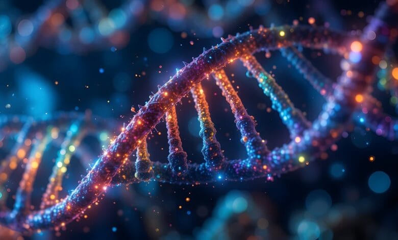
Real-time DNA damage imaging is transforming how scientists study genome maintenance. Researchers at Utrecht University have created a fluorescent live-cell DNA sensor that enables continuous visualization of DNA damage formation and repair, providing unprecedented insight into cellular responses and repair timelines.
The quest to visualize DNA in action
For decades, understanding DNA damage and repair has relied on static snapshots: cells were frozen, fixed, and analyzed at discrete time points. This approach, while informative, obscures the dynamic nature of genome maintenance.
The new sensor uses a fluorescent tag that selectively binds to damaged DNA sites. These sites glow when damage occurs and fade as repair progresses, allowing researchers to follow the entire repair sequence in living cells. Importantly, the sensor is gentle, reversible, and minimally disruptive to the cell’s natural repair machinery, addressing a key limitation of previous methods.
How the sensor works
The sensor operates through a modular design:
- Damage recognition: It binds specifically to a molecular marker that appears only on damaged DNA.
- Fluorescent tagging: The bound marker fluoresces, highlighting damage locations in real-time.
- Dynamic observation: The fluorescence fades as repair proteins complete restoration, providing a live, continuous readout of repair events.
This approach has been validated not only in cultured cells but also in living organisms, such as Caenorhabditis elegans. The team successfully monitored programmed DNA breaks during development, demonstrating the sensor’s versatility beyond cell cultures.
Seeing DNA repair unfold in a living organism was a pivotal moment. It confirmed our sensor works in real biological contexts, not just artificial lab conditions.
Illuminating the repair timeline
Real-time imaging revealed several surprising details about DNA repair:
- Rapid formation of damage sites: Some DNA breaks appear and recruit repair proteins within minutes.
- Protein recruitment dynamics: Different repair proteins arrive and leave at specific stages, forming coordinated repair hubs.
- Restoration efficiency: In many cases, the DNA returns to its intact state quickly, but slower repair in some genomic regions highlights potential vulnerabilities.
These observations underscore the importance of spatiotemporal dynamics in understanding genome integrity, revealing patterns invisible to traditional snapshot-based methods.
Mapping damage across the genome
The sensor’s modular design allows researchers to pinpoint where damage occurs in the genome. By combining this with fluorescent tagging of specific repair proteins, scientists can track protein recruitment to individual sites. Furthermore, the sensor enables repositioning of damaged DNA inside the nucleus, providing insights into how genomic location influences repair efficiency.
Not all DNA is equal. Location within the nucleus can dictate how fast or slow repair happens, and our sensor helps us see that in real-time.
Applications in cancer and aging research
Genome instability is central to cancer and age-related diseases. Real-time DNA damage imaging allows researchers to:
- Observe how chemotherapeutic drugs induce DNA damage and how efficiently cells repair it.
- Track repair in aging cells to understand accumulation of genomic errors over time.
- Identify differences between normal and cancerous cells in repair pathways.
The sensor could guide more precise drug testing and inform therapies targeting repair mechanisms, offering a high-resolution tool to improve patient safety.
By watching repair unfold live, we can identify potential drug effects before they cause harm—a proactive step in personalized medicine.
Early insights into mutagen exposure
Mutagens such as UV light or chemical agents can induce DNA damage. Traditional studies provide average effects, but real-time imaging captures heterogeneity across cells. Researchers observed:


- Some cells repair damage almost instantaneously.
- Others show delayed repair, suggesting susceptibility to mutations.
- Dynamic interplay between multiple repair proteins varies across cell types.
These insights highlight the value of continuous monitoring for understanding how environmental exposures influence genomic stability.
While promising, the technique has limitations:
- Sensor specificity: Although highly selective, there may be rare off-target binding.
- Fluorescent phototoxicity: Prolonged imaging can stress cells, requiring careful calibration.
- Complex organism imaging: Larger tissues may require advanced microscopy to maintain resolution.
These constraints are explicitly noted by the researchers, emphasizing the need for cautious interpretation of findings.
The sensor’s flexibility opens multiple research avenues:
- Studying DNA repair in complex tissues or organoids.
- Investigating the interplay between DNA damage and transcriptional activity.
- Exploring repair mechanisms in human stem cells or primary patient samples.
Because the sensor is freely available, laboratories worldwide can rapidly adopt and adapt the technology, accelerating discoveries in genome maintenance and therapeutic research.
Prior studies have used fixed-cell imaging, comet assays, or immunofluorescence to study repair. While informative, these methods cannot capture the continuous kinetics of damage and repair. Real-time sensors thus fill a crucial gap, providing temporal resolution while preserving cellular integrity.
Ongoing debates focus on balancing imaging resolution with cellular stress, and how to generalize findings from model organisms to humans. The Utrecht sensor addresses these issues by minimizing interference and enabling observations across scales.
References:
- Jackson, S. P., & Bartek, J. (2009). The DNA-damage response in human biology and disease. Nature, 461, 1071–1078. https://doi.org/10.1038/nature08467
- Ciccia, A., & Elledge, S. J. (2010). The DNA damage response: making it safe to play with knives. Molecular Cell, 40(2), 179–204. https://doi.org/10.1016/j.molcel.2010.09.019
Disclaimer: Some content on this page may have been created or reviewed with the help of artificial intelligence tools. While every effort is made to ensure reliability, readers are advised to consult primary sources. External links and references are offered for convenience, and Honores is not liable for their content or impact.